
Open Magnetic Resonance Imaging (MRI)
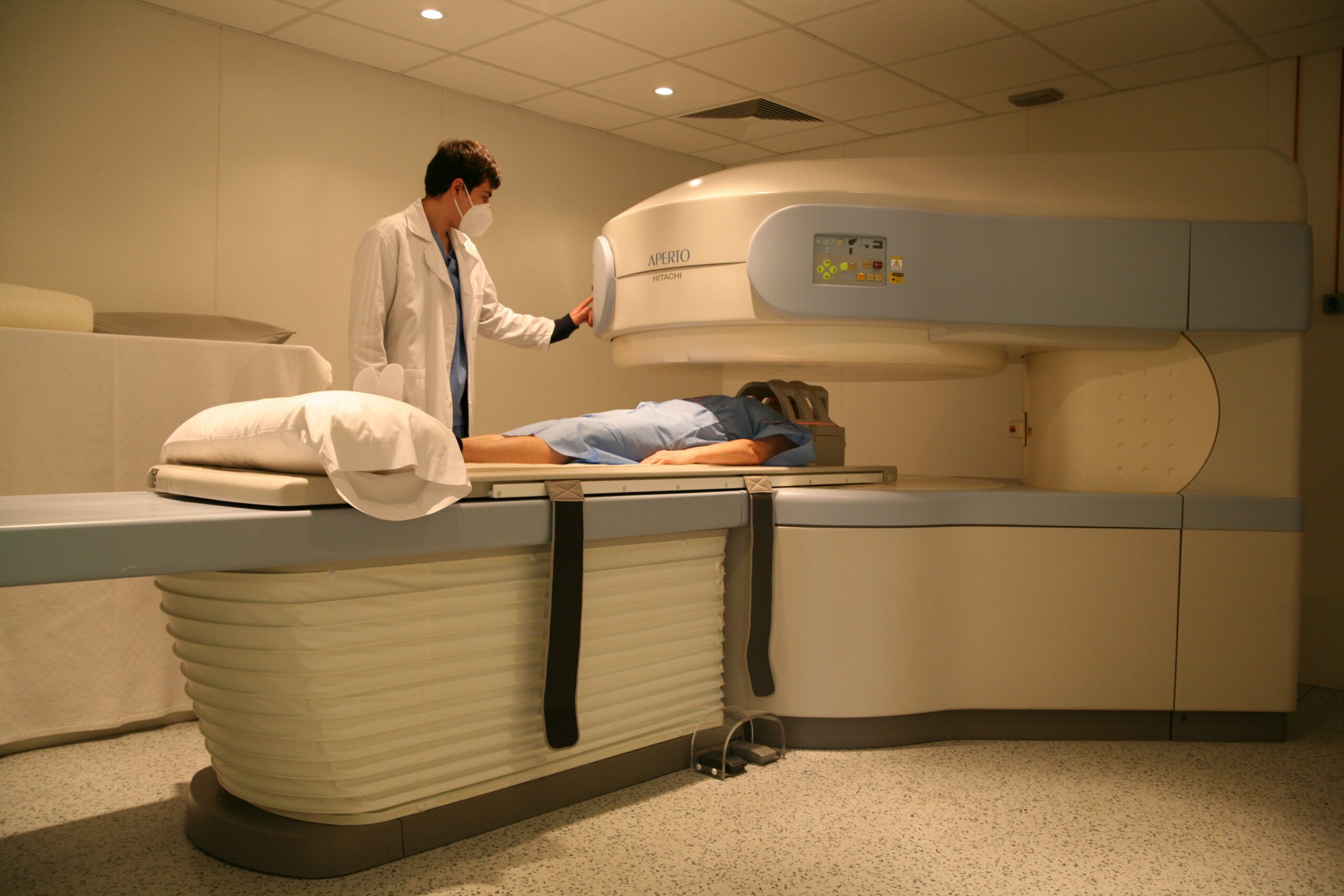
Nuclear Magnetic Resonance allows diagnostic imaging tests of the whole organism and specific tests of the structures of the central and peripheral nervous system. Open Magnetic Resonance Imaging facilitates the performance of tests in people who have difficulties to remain immobile in a closed system, such as people who suffer from claustrophobia. people suffering from claustrophobia.
-

- Un paciente se recuesta en una mesa, listo para que le hagan una resonancia magnética en un centro médico. Un profesional sanitario con bata blanca se encuentra de pie junto a la máquina de resonancia magnética, comprobando los controles. La sala está bien iluminada y equipada para realizar procedimientos de diagnóstico por imagen.
-
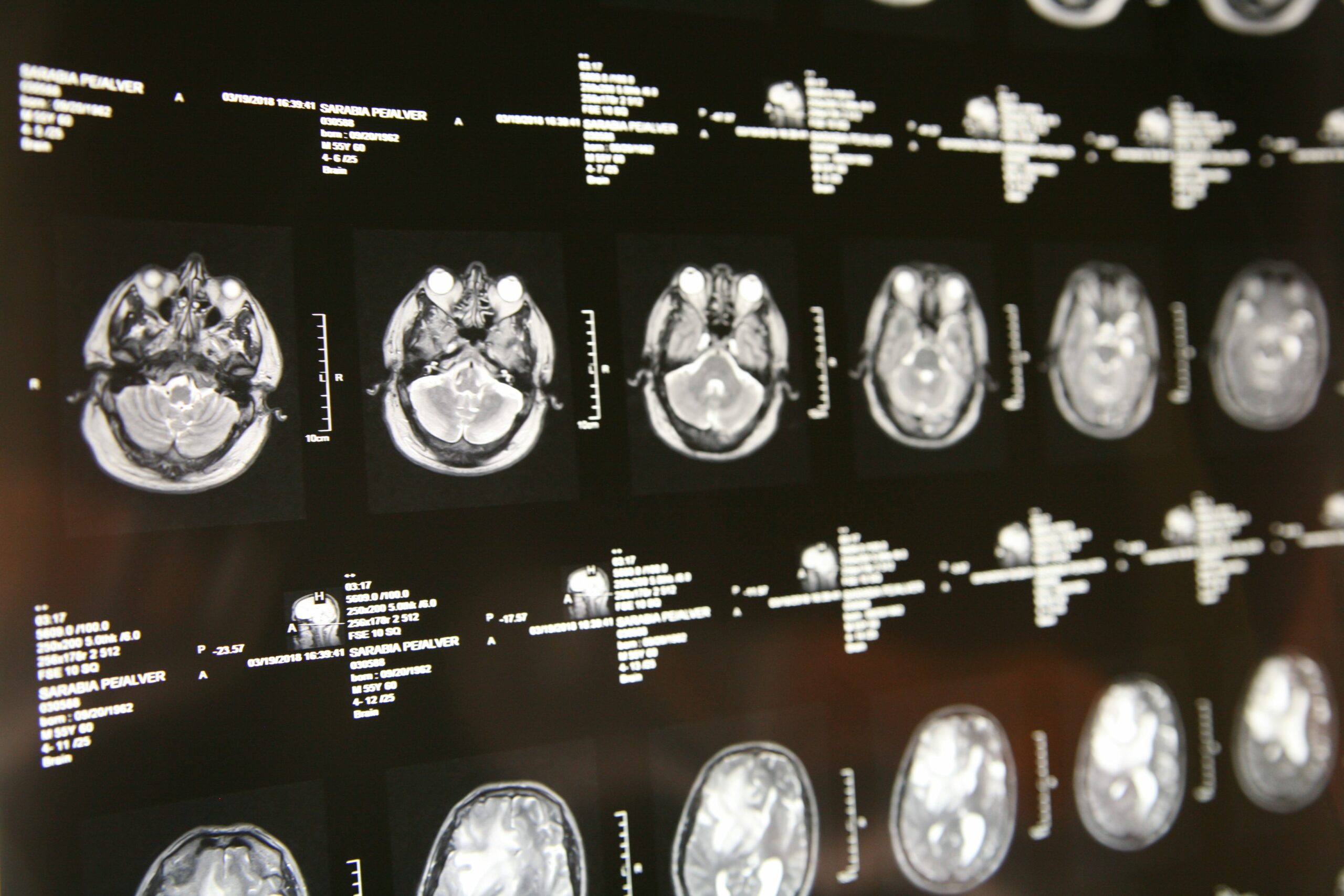
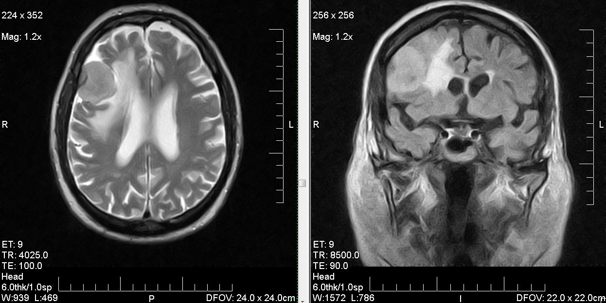
- Una serie de imágenes de resonancia magnética cerebral mostradas en un panel retroiluminado, que muestran múltiples imágenes transversales con distintos detalles de las estructuras cerebrales. Etiquetas de texto blancas y escalas de medición acompañan cada imagen.
-
- Magnetic Resonance Pathology Imaging

Computed Axial Tomography (CAT) scan
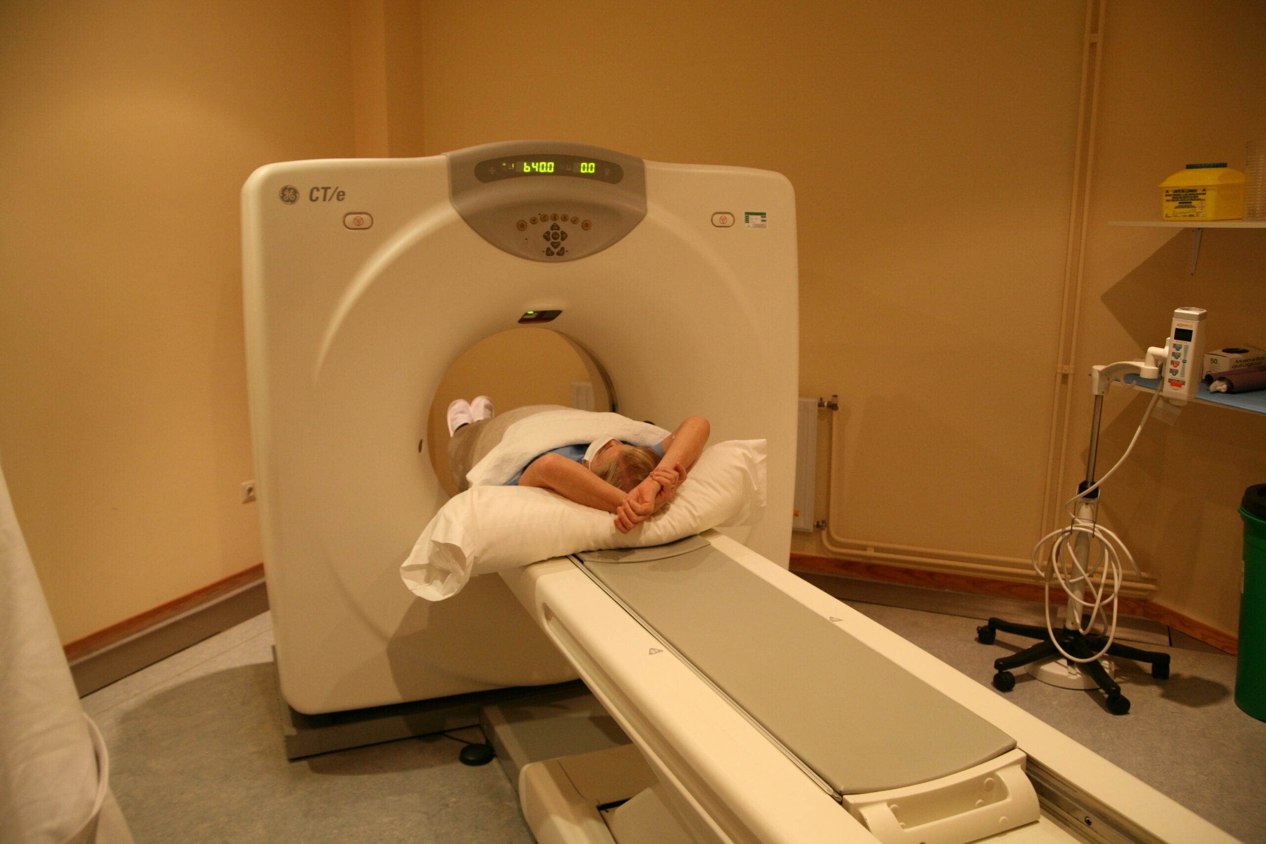
Computed Axial Tomography, also known by the acronym CT or by the name scanner, is an X-ray scan that produces images of axial slices of the body. It obtains very precise images of the interior of the organism and its different organs, allowing very rigorous diagnoses.
-
- TAC
-
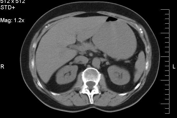
- Abdominal CT image
-
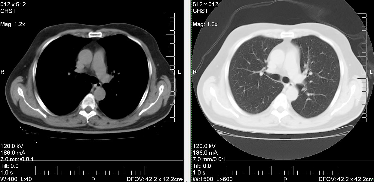
- CT lung imaging

Conventional radiology
Conventional radiology is the most common method of radiodiagnosis, and is still the most commonly used modality today. Radiological images are obtained quickly and show the anatomy of the human body in grayscale, allowing the detection and diagnosis of different pathologies.
-
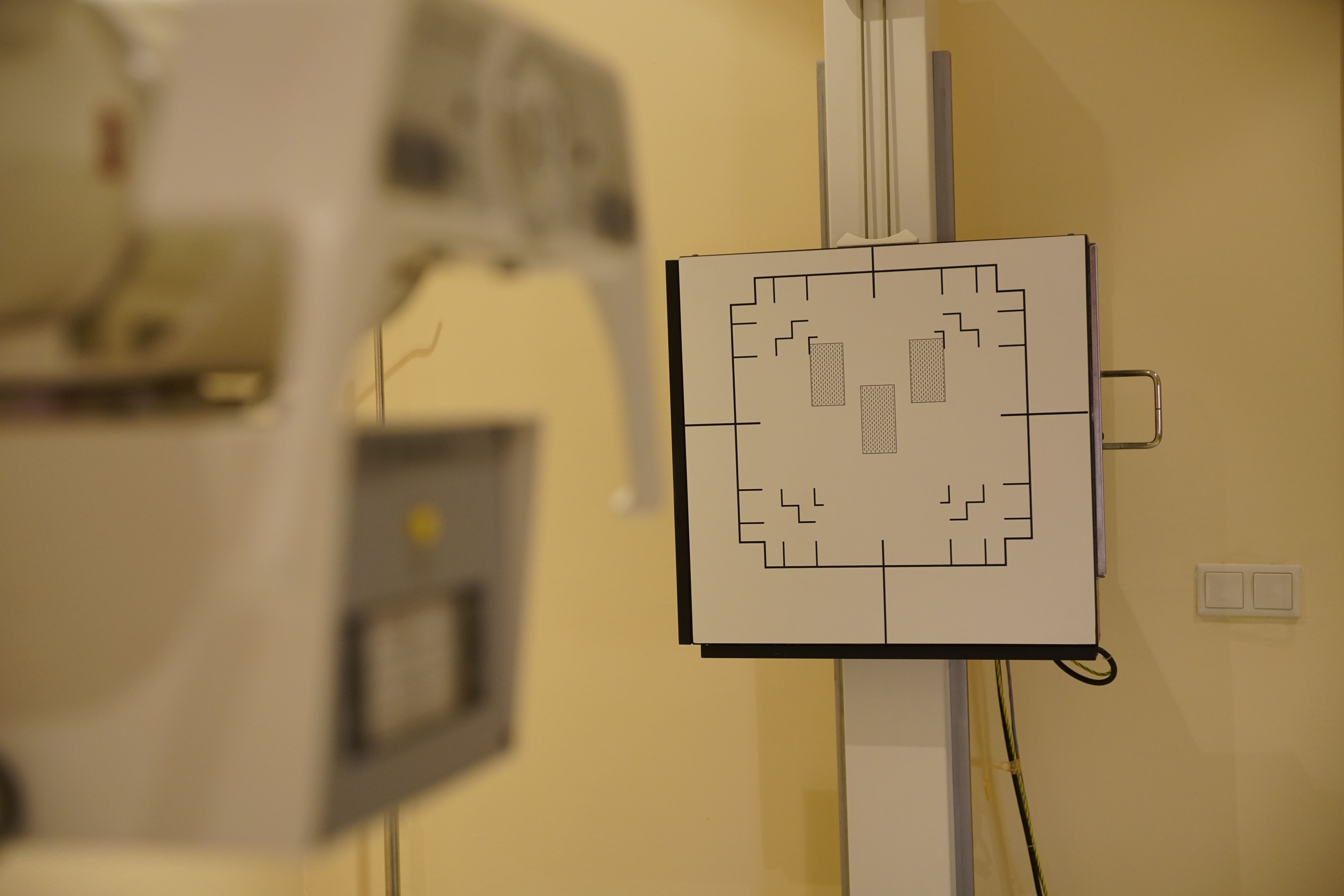
- X-Ray Generator
-
- X-ray of cast arm

-
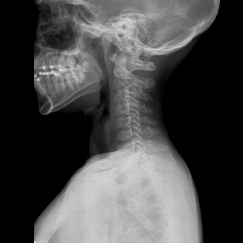
- Lateral neck x-ray

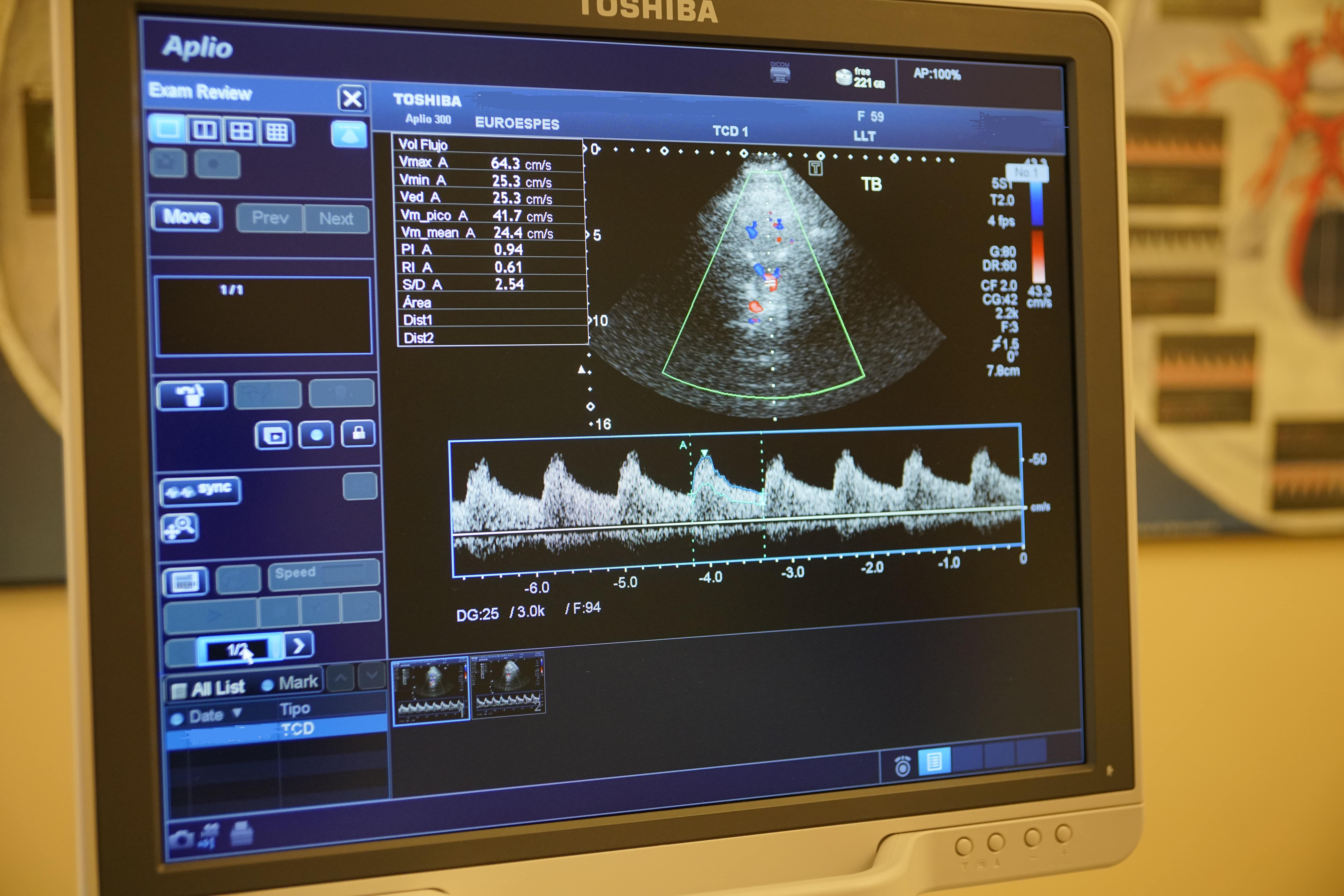
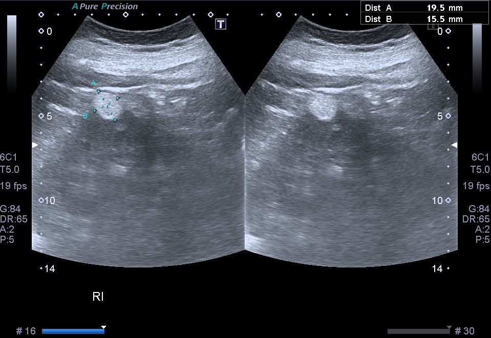
Conventional / transcranial ultrasound
Thanks to ultrasound we can provide images in the following clinical applications: pediatric, small parts, breast, abdomino-pelvic, urological, gynecological, musculoskeletal, cardiac (adult and pediatric), echo-Doppler, peripheral vascular... Transcranial Doppler ultrasonography allows to evaluate hemodynamic parameters in the cerebral circulation.
-
- Ultrasound scanner

-
- Doppler imaging
-
- Ultrasound image with pathology

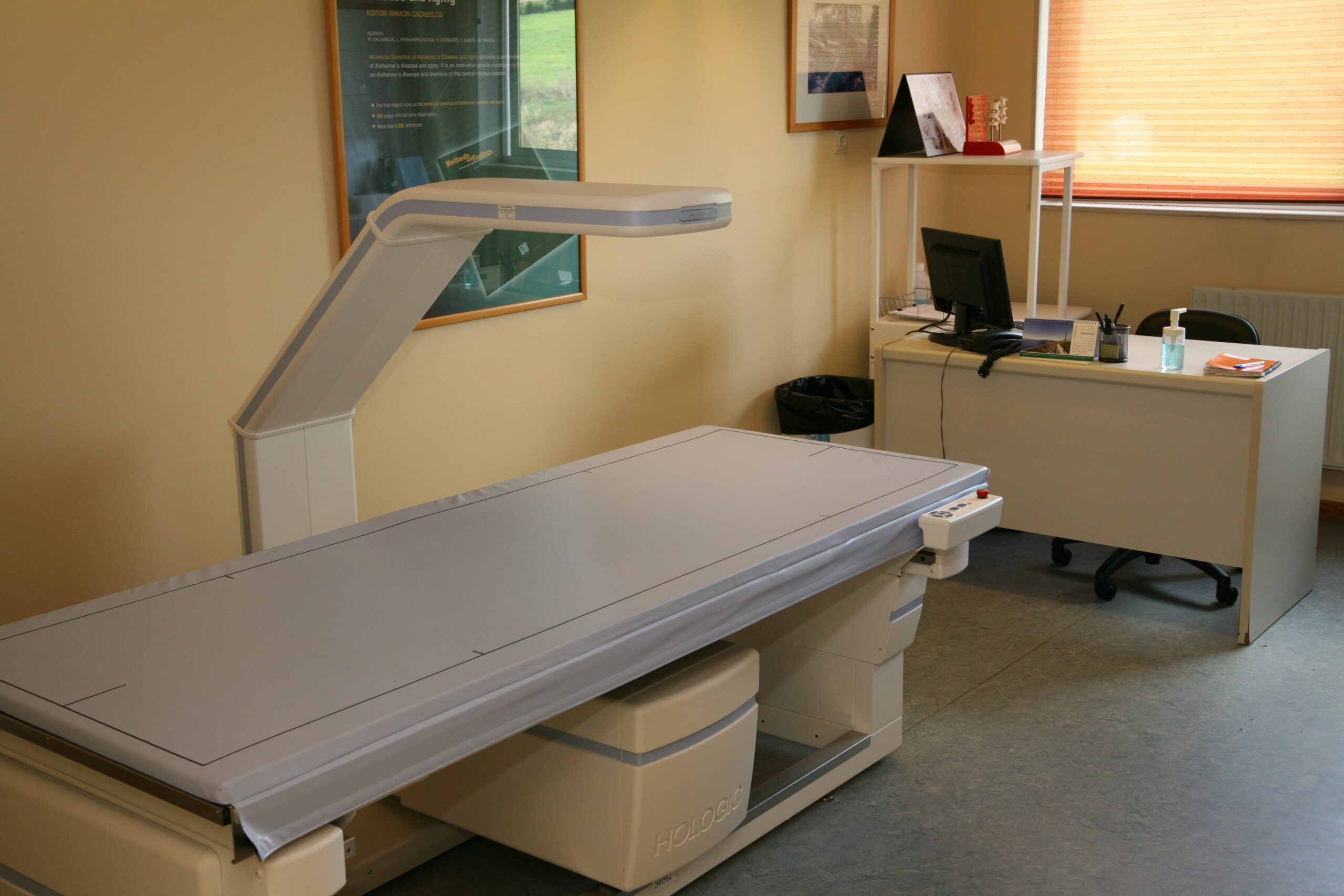
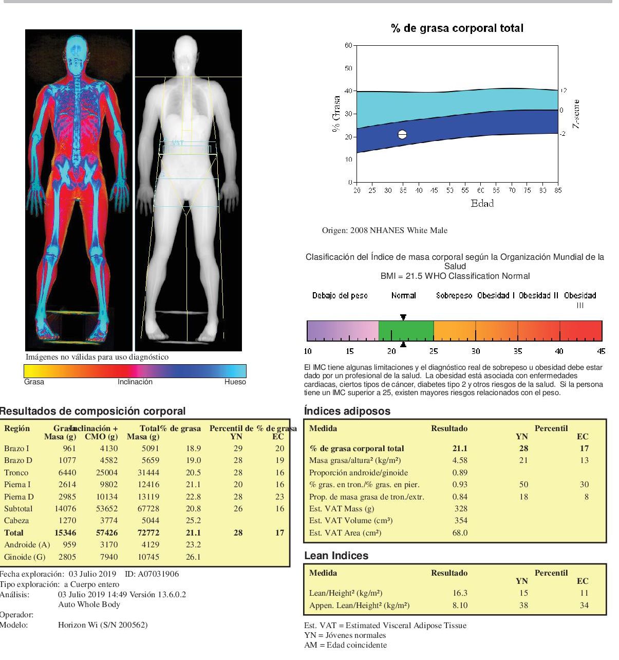
Densitometry
Used for the diagnosis of bone pathologies, such as osteoporosis and osteopenia; to assess the risk of fractures; to study the quantity, distribution and osteoporosis and osteopenia; to assess the risk of fractures; to study the amount, distribution and ratio of bone, fat and lean (muscle) tissue in different body regions; to calculate BMI (body mass index).
-
- Densitometer
-
- Thermography report
-
- Densitometry report
